
Blog


Accelerating Biomedical Innovations with FRED
In:
Created:
24 Jul 2024
Introduction
From non-invasive procedures to ultra-sensitive diagnostic instrumentation, photonic devices play an indispensable role in today’s biomedical industry. Timely design and delivery to market of these new technologies have been made possible with the aid of sophisticated software tools and experienced optical engineers. Photon Engineering firmly believes that its optical engineering product, FRED, can help accelerate the pace of innovation in the biomedical community. FRED combines an intuitive graphical user interface with a powerful computational engine capable of satisfying the most demanding requirements. The relevance of FRED to the biomedical industry can best be expressed by presenting several familiar yet innovative applications such as a gonioscope, laser-induced fluorescence in a capillary, and a human skin model.
Biomedical Optics Applications
Gonioscopy Lens
Monitoring the iridocorneal angle (the angle between the iris and the internal surface of the cornea) is crucial for diagnosing and treating glaucoma. A gonioscope measures this angle by illuminating the eye and collecting reflected light. To simulate the gonioscope, an accurate human eye model is required. A model based on relevant references is available in the Samples folder of FRED, including all essential elements: front and rear surfaces of the cornea, iris, eye lens, and aqueous humor. To modify any aspect of the eye model, simply double-click each element to open a multi-tab dialog box.

Figure 1. FRED model: anterior portion of the human eye.
Next, model the gonioscopy lens. FRED allows the import of lenses designed in CodeV, Zemax, or Oslo. FRED maintains a local coordinate system for each surface, allowing precise positioning of the goniolens to the cornea. In practice, the lens is coupled to the cornea using index-matching fluid. FRED’s unique “gluing” feature facilitates easy insertion of this layer. To contact the surfaces, enter edit mode for the rear surface of the goniolens, click the Glue tab, and select the Cornea outer surface as the surface to be glued to. Finally, select the material for gluing.

Figure 2. “Gluing” the goniolens to the cornea.
Create the light source for illumination by generating a Detailed Optical Source.

Figure 3. Detailed Optical Source dialog box. The Positions/Directions tab specifies source dimensions, number of rays, and angular emission properties. The Wavelengths tab provides options for spectral content. The Power tab sets total source power and specifies any spatial apodization.
The completed and raytraced model is shown below. FRED allows assigning colors to rays under specific conditions: reflection, transmission, scattering, or diffraction. In this example, rays scattering from the cornea rear surface are green, and those scattering from the iris are red.

Figure 4. Goniolens model with raytrace paths. Ray colors indicate specific surface intersections.
Spot diagrams at the lens focus for rays scattered from the iris and the cornea are shown below. The left and right plots contrast the difference between flat and curved irises. As expected, the image shears with the curved iris in the left chart.

Figure 5. Spot diagrams using planar and curved irises.
Laser-Induced Fluorescence – Capillary Electrophoresis
Capillary electrophoresis is a technique used in genetic analysis and protein characterization. A collimated laser beam is focused into a glass capillary column where material flows under an electric potential. Particles passing through the illuminated volume fluoresce with a characteristic spectrum.
Below, a collimated rayset representing a UV laser beam is focused by an objective lens into a glass capillary filled with fluid. The mirror at the top right enlarges the illuminated volume by reflecting unused light back into the capillary at a slightly different trajectory, increasing the fluorescent signal. Optics perpendicular to the illumination path collect fluorescent light for analysis.

Figure 6. FRED model of capillary electrophoresis system with collection optics.
FRED can simulate fluorescence through its scattering library. A scripted scatter model can reassign ray wavelengths by interpreting an emission curve in terms of probability. Here, Rhodamine 6G, a widely used organic dye, is used. The spectrum is digitized in FRED and included in a Scripted Scatter Model.
To save simulation time, only scattered rays reaching the detector should be traced. The Importance Sampling feature in FRED serves this function. Select the fluorescing object, click the Scatter tab, assign the Scripted Scatter property of fluorescence, and edit the Scatter Direction Region(s) to be “Toward an Entity,” selecting the detector surface as this entity.
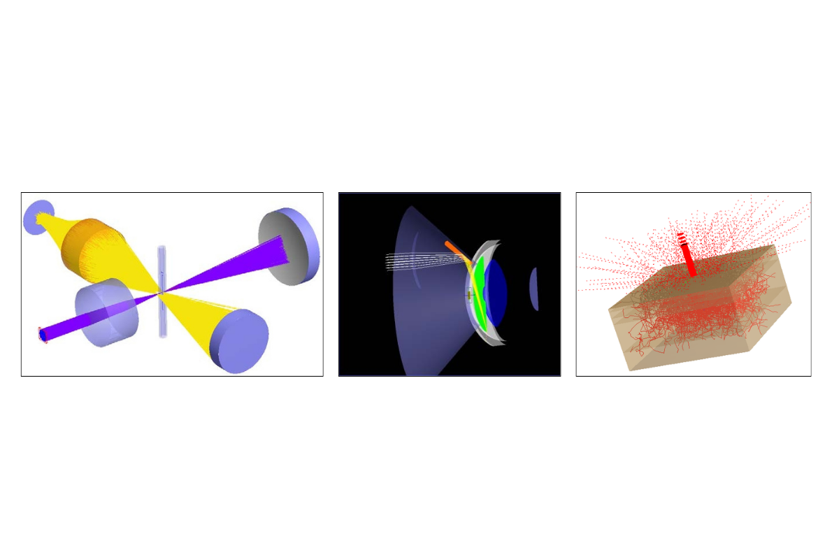
A graphical representation of the complete simulation is shown below. Purple represents the illuminating path, while orange maps the fluorescence.

Figure 7. Capillary Electrophoresis simulation with illumination and fluorescence paths.
Human Skin Model
Human skin models are valuable for designing non-invasive diagnostic devices and dermatological instruments. FRED offers the Henyey-Greenstein volume scatter model, recognized as representative of scattering in human tissue. This model is applied through a material definition in FRED. Once defined, it can be assigned to entities in the model by drag-and-drop.

Figure 8. (Left) Defining the Henyey-Greenstein volume scatter model for a new material: top – scattering properties; bottom–absorption properties. (Right) Sample raytrace of a human skin model in FRED.
As demonstrated in these biomedical optical device examples, FRED possesses critical and visually dynamic capabilities for modeling, analysis, and graphical display. If you have any questions regarding FRED’s ability to model and analyze your biomedical optical system, feel free to contact us by phone or email.
___________________________________________________________________________________________________________________________________________________________________________________
This blog post was created based on the information provided by Photon Engineering, a partner of CBS Europe.